Lamina dura
- Dr Chandan Dolare
- Jan 19, 2020
- 4 min read
The name lamina dura is applied to the thin layer of dense, cortical bone which lines the root socket of the alveolar process. It is visualized as a thin opacity since it results due to a layer of dense bone.
The thin hypodensity/ radiolucency present along the tooth side denotes the periodontal ligament space and cancellous bone lies on the opposite side of the lamina dura.
The lamina dura can be demarcated around a recent extraction socket.


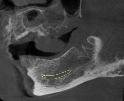
Coronal image showing socket with 12 and sagittal image showing socket with 38. Lamina dura is demarcated around the alveolar socket.
CBCT is considered better than IOPA/ Periapical radiographs and Panoramic radiographs for detection of lamina dura and PDL space.(1) CBCT can detect bone defects of the cancellous bone and cortical bone separately.
The CBCT volumetric data set consists of isotropic voxels. Majority of the software used for assessment allows multiplanar reformation (MPR) which can generate non-orthogonal 2D images. The MPR modes include Oblique, Curved planar reformation, which can be used to evaluate focused anatomic areas under investigation.
Thus, the differences in the shape and contour of the root/s do not affect the width and/or density of the lamina dura in images derived from multiplanar reconstruction in CBCT as there is no superimposition of anatomical structures or distortion due to insufficient thickness.
The axial, coronal and sagittal reconstructed images without superimposition, magnification and distortion can be used to detect involvement of lamina dura and PDL space in periodontal lesions. Thus lesions showing areas with furcation involvement; buccal and lingual/ palatal bone discontinuity; and intrabony defects can be detected using multiplanar reconstruction in CBCT. (2)
Similarly the complex relationship and borders between teeth and their anatomic structures such as the maxillary sinus and mandibular canal, mental canal and foramen are clearly visualized. This helps to eliminate errors caused due to superimposition in 2D radiography.
Factors to be considered or evaluated in the absence of lamina dura
Integrity of the cortical lining of lamina dura
In majority of cases, the lamina dura can be demarcated from the crestal area along the root length and into the furcation area for a normal tooth.
Previous radiographs
Evaluate earlier images to check if the lamina dura has changed.
Normal anatomic variations
Certain radiolucencies may be noted entering the lamina dura, which could be suggestive of anatomic vascular canals/ foramens.

Coronal images showing nutrient canals i.r.t
socket of 43 and 33
External root resorption
In cases of external root resorption, loss of dentinal structure is noted at the affected area. However, lamina dura usually stays intact and can be demarcated


Cross sectional and axial images showing resorption i.r.t the outer aspect of the root of 21. Lamina dura can be demarcated along the resorbed root surface.
Diseases causing discontinuity or loss of lamina dura
Discontinuity in lamina dura is indicative of abnormality, usually suggestive of disease. Most common causes for the absence of lamina dura are periapical inflammatory lesions and periodontal disease.
The common causes for localized loss of lamina dura are – apical abscess, periapical granuloma, periapical cyst, periapical cementosseous dysplasia, osteomyelitis.
Periapical inflammatory disease

Periapical abscess
Sagittal image showing localized osteolytic process and loss of lamina dura i.r.t mesial and distal roots of 38.

Periapical granuloma
Sagittal images showing ovoid, well demarcated, radiolucent lesion with sclerotic periphery. Loss of lamina dura is noted i.r.t palatal root of 16 along with displacement of the sinus floor

Periapical cyst
Sagittal image showing unilocular, cystic lesion with well defined periphery i.r.t distal root of carious 36. Periapical osteolysis noted with mesial root. Loss of lamina dura is noted with both roots of 36 and mesial root of 37 (yellow marker).
Periodontal disease
A discontinuity in the lamina dura and a wedge-shaped radiolucent area along the mesial and/or distal aspect of the periodontal ligament space is the earliest sign of periodontal disease.

Sagittal images for right maxilla showing furcation bone loss and severe distal bone loss noted i.r.t 16. Periradicular periodontitis is noted along with loss of lamina dura.

Periodontal disease and Infrabony defect : Coronal and axial images showing infrabony defect i.r.t 42 along with discontinuity in lamina dura. Widening of periodontal ligament space is also noted i.r.t 42,43.
Focal and Periapical cementosseous dysplasia

Sagittal image showing loss of lamina dura i.r.t the distal root of 36. Bulbous apical 3rd region noted with the distal root of 36.
Ameloblastoma

Sagittal CBCT image shows multilocular expansile lesion with right body-ramus of mandible showing defect with crestal bone and alveolus i.r.t 48 region. Loss of lamina dura is noted i.r.t the roots of 47 along with resorption. Apical resorption and loss of lamina dura is noted with distal root of 46.
References :-
Nimish Prakash, Karjodkar FR, Sansare K, Sonawane HV, Bansal N, Arwade R. Visibility of lamina dura and periodontal space on periapical radiographs and its comparison with cone beam computed tomography. Contemp Clin Dent 2015; 6: 21–5. doi: https://doi.org/10.4103/0976-237x.149286 [PMC free article] [PubMed] [Google Scholar]
Acar B, Kamburoğlu K. Use of cone beam computed tomography in periodontology. World J Radiol. 2014;6(5):139–147. doi:10.4329/wjr.v6.i5.139
Oral Radiology : Principles and interpretation 5th edition White & Pharoah
Clinical Periodontology 9th edition Newman, Takei & Carranza
Color atlas of Anatomy 3rd edition Rohen and Yokochi
Differential diagnosis of Oral and Maxillofacial lesions 5th edition, Wood and Goaz
Normal radiographic appearances and variations – HM Worth
